

| Back | Interactive CS16 Embryo Model |
|
|
Key to anatomical regions
|
| Back | Movies of CS16 embryos |
The movies below show volume renders of OPT models. No staining has been used on any of the specimens and the images were captured using autofluorescence with a FITC filter.
| N346 46XX |
N376 46XX |
N426 46XX |
| N519 46XY |
N709 46XX |
N1323 46XX |
The movies below show volume renders of High Resolution Episcopic Microscopy (HREM) models. Other HREM models can be found here.
|
1716 46XY |
1815 46XY
|
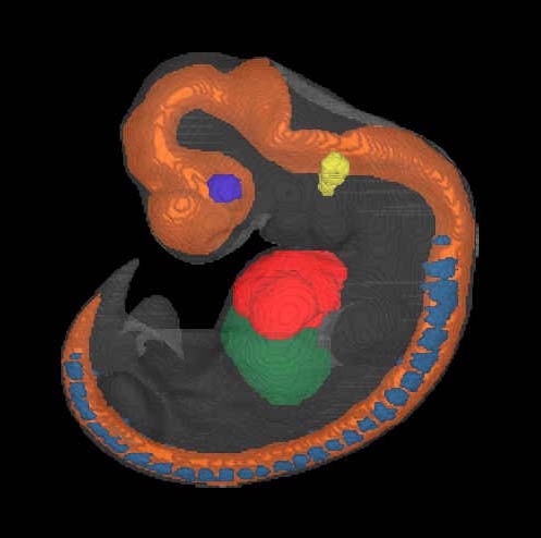
| Back | Painted Anatomical Regions |
The movies below show anatomical domains defined in the N346 model. The domains have been manually defined in the OPT model using specialised software and then visualised using commercial rendering packages.
The various domains are: neural tube - orange; heart - red; liver - dark green; optic vesicle - dark blue; otic vesicle - yellow; dorsal root ganglia - cyan.
| N346 46XX |
| Back | Carnegie Stage 16 Definition |
| Age is approximately 38 postovulatory days Length is approximately 7 - 12 mm External Features Include: retinal pigment becoming visible externally; the thigh, leg and foot are becoming distinguishable; pharyngeal arch 2 is more massive and more conspicuous (whereas arch 3 is receding from the surface); auricular hillocks are beginning to appear. O'Rahilly R and Muller F [1987] Developmental Stages in Human Embryos. Carnegie Institute, Washington, Publication no. 637, and O'Rahilly R, Müller F (2010) Developmental stages in human embryos: revised and new measurements. Cells Tissues Organs. 192:73-84. PMID: 20185898. |