

| Back | Interactive CS17 Embryo Model - sample 365 |
|
|
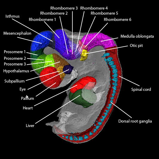
Key to anatomical regions
|
| Back | Movies of CS17 embryos |
The movies below show volume renders of OPT models. No staining has been used on any of the specimens and the images were captured using autofluorescence with a FITC filter.
| N363 46XX |
N365 46XX |
N366 46XY |
| N471 46XX |
N478 46XY |
N486 |
| N512 46XY |
N522 46XX |
N544 46XY |
| N522 46XX |
N571 46XX |
The movie below shows a volume render of a High Resolution Episcopic Microscopy (HREM) model. Other HREM models can be found here.
|
1699 46XY
|
| Back | Painted Anatomical Regions |
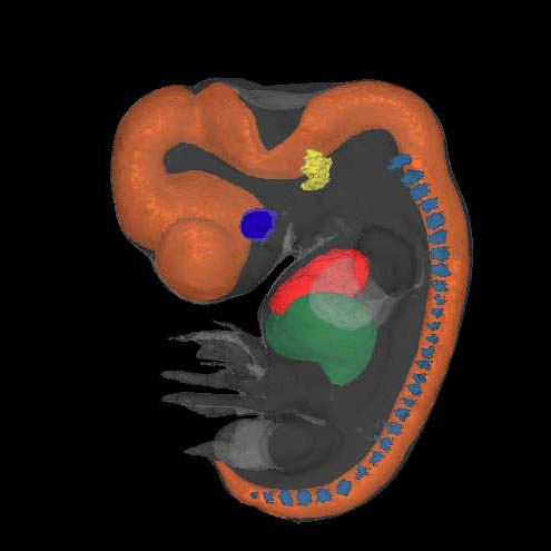
The movies below show anatomical domains defined in the N365 model. The domains have been manually defined in the OPT model using specialised software and then visualised using commercial rendering packages.
The various domains are: heart - red; liver - dark green; eye - dark blue; otic vesicle - yellow; dorsal root ganglia - cyan. The CNS domains have been further subdivided as follows: pallium - red, subpallium - orange, hypothalamus - light brown, prosomere 3 - bright green, prosomere 2 - green, prosomere 1 - dark green; mesencephalon - blue; isthmus - dark purple, metencephalon (rhombomeres 1-6) - purples/pinks; myelencephalon (rhombomeres 7-11) - magenta; spinal cord - maroon.
The top panel is a surface render of the 3D anatomical regions, and the bottom panel shows the painted N365 model being digitally sectioned through the z-plane.
 |
Immunohistochemistry
The movies below show rendered representations of immunohistochemistry data from sections through the head of N365 which have been digitised, thresholded out and placed back onto the OPT model of N365.
PAX6 |
GAP43 |
Beta-tubulin |
PAX6: The staining is shown in red. For reference the eyes have been shown in dark grey, and the PAX6-negative regions of the neural tube are shown in light grey.
GAP43: Here the individual cranial nerves have defined and painted as follows: Olfactory (I) - light blue, Oculomotor (III) - yellow/green, Trochlear (IV) - blue/green, Trigeminal (V) - violet, Abducens (VI) - deep orange, Facial (VII) - pink, Vestibulocochlear (VIII) - white, Glossopharyngeal (IX) - light orange, Vagus (X) - red, Accessory (XI) - bright green, Hypoglossal (XII) - yellow and spinal nerves bright blue. Only one hemisphere is shown to allow visualisation of the nerves from both sides. For reference the eye is painted dark grey and the neural tube light grey.
B Tubulin: The strong staining is shown in red while the orange represents the weaker staining. The eyes have been shown in dark blue for reference purposes.
| Back | Carnegie Stage 17 Definition |
Age is approximately 40 postovulatory days
Length is approximately 10 - 14mm
External Features Include: the hand plate exhibits definite digital rays; the foot has acquired a rounded digital plate; nasofrontal grooves are distinct.
O'Rahilly R and Muller F [1987] Developmental Stages in Human Embryos. Carnegie Institute, Washington, Publication no. 637, and O'Rahilly R, Müller F (2010) Developmental stages in human embryos: revised and new measurements. Cells Tissues Organs. 192:73-84. PMID: 20185898.