

| Back | Interactive CS20 Embryo Model - sample 913 |
The model is high resolution. Please allow time for it to load.
|
|
Key to anatomical regions
|
| Back | Movies of CS20 embryos |
The movies below show volume renders of OPT models. Images of unstained embryos were captured using autofluorescence with a FITC filter.
| N382 46XX |
N529 46XY |
The movies below show volume renders of High Resolution Episcopic Microscopy (HREM) models. Other HREM models can be found here.
|
N913 46XX
|
1645 46XX
|
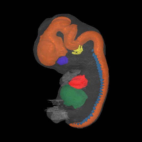
| Back | Painted Anatomical Regions |
The movie below shows anatomical domains defined in the N382 model. The domains have been manually defined in the OPT model using specialised software and then visualised using commercial rendering packages.
The various domains are: neural tube - orange; heart - red; liver - dark green; optic vesicle - dark blue; otic vesicle - yellow; dorsal root ganglia - cyan.
| N382 46XX |
| Back | Carnegie Stage 20 Definition |
Age is approximately 49 postovulatory days
Length is approximately 18 - 23mm
External Features Include: the upper limbs have become slightly bent at the elbows; hands have short, stubby fingers and are still far apart, a delicate fringe-like vascular plexus appears in the superficial tissue of the head and the edge of the plexus is approximately halfway between ear-eye level and the vertex of the head.
O'Rahilly R and Muller F [1987] Developmental Stages in Human Embryos. Carnegie Institute, Washington, Publication no. 637, and O'Rahilly R, Müller F (2010) Developmental stages in human embryos: revised and new measurements. Cells Tissues Organs. 192:73-84. PMID: 20185898.