

| Back | Interactive CS14 Embryo Model - sample A386 |
|
|
Key to anatomical regions
|
| Back | Movies of CS14 embryos |
The movies below show volume renders of OPT models. No staining has been used on any of the specimens and the images were captured using autofluorescence with a FITC filter.
| A386 46XX |
N420 46XY | N518 46XX |
| N560 46XY |
N578 46XY |
N939 46XY |
The movies below show volume renders of High Resolution Episcopic Microscopy (HREM) models. Other HREM models can be found here.
|
N925 46XX |
N933 46XX |
| Back | Painted Anatomical Regions |
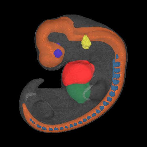
The movies below show anatomical domains defined in the A386 and N420 models. The domains have been manually defined in the OPT models using specialised software and then visualised using commercial rendering packages.
The various domains are: neural tube - orange; heart - red; liver - dark green; optic vesicle - dark blue; otic vesicle - yellow; dorsal root ganglia - cyan.
| A386 46XX |
N420 46XY |
| Back | Carnegie Stage 14 Definition |
| Age is approximately 34 postovulatory days Length is approximately 5 - 8 mm External Features Include: elongated and tapering upper limb buds, invagination of the lens disc but with open lens pit. Internally, future cerebral hemispheres and cerebellar plates begin to be visible. O'Rahilly R and Muller F [1987] Developmental Stages in Human Embryos. Carnegie Institute, Washington, Publication no. 637, and O'Rahilly R, Müller F (2010) Developmental stages in human embryos: revised and new measurements. Cells Tissues Organs. 192:73-84. PMID: 20185898. |