

| Back | Interactive CS19 Embryo Model - sample 454 |
The model is high resolution. Please allow time for it to load.
|
|
Key to anatomical regions
|
| Back | Movies of CS19 embryos |
The movies below show volume renders of OPT models. No staining has been used on any of the specimens and the images were captured using autofluorescence with a FITC filter.
| N454 46XY |
N534 46XY |
| N337 46XY |
N344 46XX |
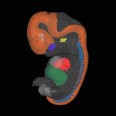
| Back | Painted Anatomical Regions |
The movies in the left and centre panels are surface renders of neuroanatomical domains. These have been manually defined in the N454 CS19 OPT model using specialised software and then visualised using commercial rendering packages.
N454 46XY |
N454 46XY |
N454 46XY |
Left: The domains are neural tube - orange; heart - red; liver - green; eye - dark blue; otic vesicle - yellow and dorsal root ganglia - light blue. Centre: The domains are eye - light grey; pallium - red, subpallium & hypothalamus - orange; prosomere 3 - yellow, prosomere 2 - bright green, prosomere 1 - dark green, mesencephalon - blue; metencephalon - purple; myelencephalon - pink; spinal cord - maroon. The movie on the right shows the same N454 model and painted anatomical domains being digitally sectioned through the z-plane. The various domains are: heart - red; liver - dark green; eye - dark blue; otic vesicle - yellow; dorsal root ganglia - cyan. The CNS domains have been further subdivided as follows: pallium - red, subpallium - orange, hypothalamus - light brown, prosomere 3 - bright green, prosomere 2 - green, prosomere 1 - dark green; mesencephalon - blue; isthmus - dark purple, metencephalon (rhombomeres 1-6) - purples/pinks; myelencephalon (rhombomeres 7-11) - magenta; spinal cord - maroon. |
| Back | Carnegie Stage 19 Definition |
Age is approximately 46 postovulatory days
Length is approximately 15 - 21 mm
External Features Include: the trunk has begun to straighten and elongate slightly, with the result that the head no longer forms a right angle with the line of the back of the specimen, the limbs extend nearly directly forward; the toe rays are more prominent, but interdigital notches have not yet appeared in the rim of the footplate.
O'Rahilly R and Muller F [1987] Developmental Stages in Human Embryos. Carnegie Institute, Washington, Publication no. 637, and O'Rahilly R, Müller F (2010) Developmental stages in human embryos: revised and new measurements. Cells Tissues Organs. 192:73-84. PMID: 20185898.