

| Back | Interactive CS23 Embryo Model - sample 14685 |
The model is high resolution. Please allow time for it to load.
|
|
Key to anatomical regions
|
| Back | Movies of CS23 embryos |
The movie below shows a micro-CT scan of a CS23 embryo. Sample staining and micro-CT was performed by Newcastle |
|
14685, male.
|
The movies below show volume renders of OPT models.
Images of autofluorescence from unstained samples were captured using a FITC filter.
| N336 46XY |
N448 46XY |
| Back | Painted Anatomical Regions |
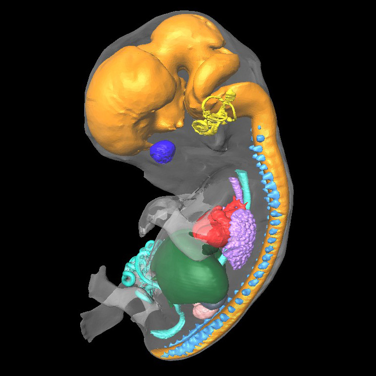
The movies below show anatomical domains defined in CS23 models.
The domains have been manually defined using specialised software and then visualised using commercial rendering packages.
14685 domains: neural tube - orange; eye - dark blue; otic vesicle - yellow; dorsal root ganglia - light blue; heart - red; liver - dark green;
gastrointestinal tract - cyan; lungs and trachea - lilac; adrenal - dark grey; kidney - pale pink.
| 14685, male |
N448 domains: neural tube - orange; eye - dark blue; otic vesicle - yellow; dorsal root ganglia - cyan.
| N448 46XY |
| Back | Carnegie Stage 23 Definition |
Age is approximately 56 postovulatory days
Length is approximately 23 - 32mm
The eyelids are starting to fuse at margins. The superficial vascular plexus is rapidly approaching the vertex of the head. The limbs have increased markedly in length and show more advanced differentiation of their subdivisions.
O'Rahilly R and Muller F [1987] Developmental Stages in Human Embryos. Carnegie Institute, Washington, Publication no. 637, and O'Rahilly R, Müller F (2010) Developmental stages in human embryos: revised and new measurements. Cells Tissues Organs. 192:73-84. PMID: 20185898.