

| Back | eHistology Viewer |
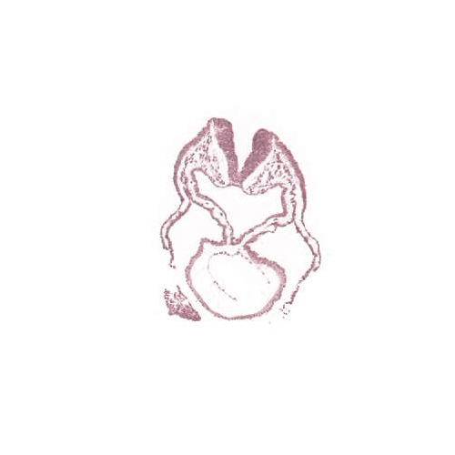
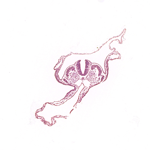
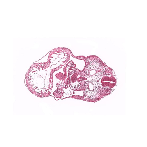
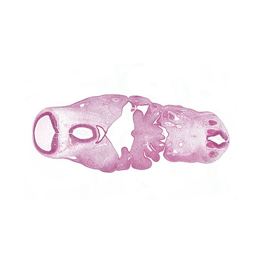
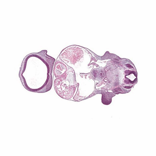
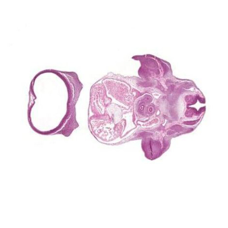
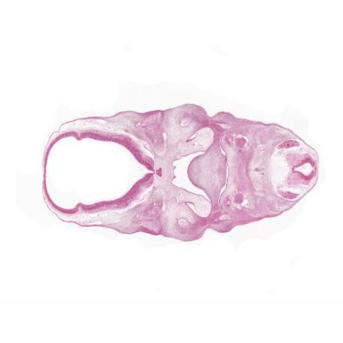
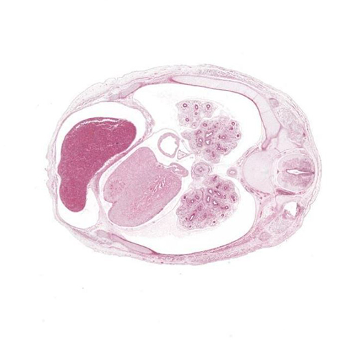
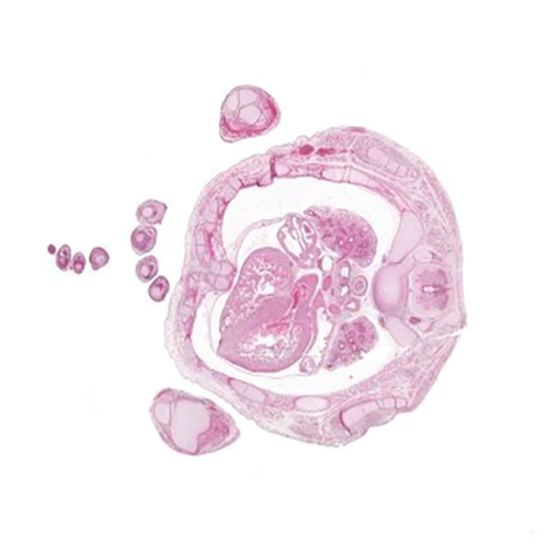
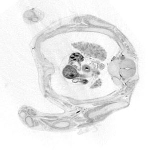
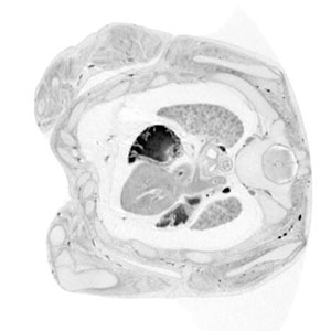
Below are sets of transverse, sagittal and frontal sections from CS10 to 14PCW.
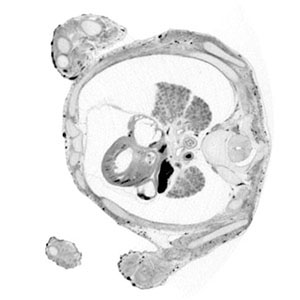
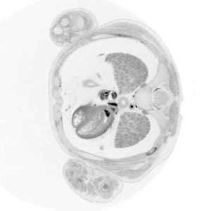
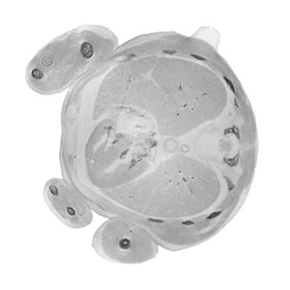
H&E sections, scanned at X20 or X40 are shown for the embryonic stages, while microCT images are shown for the fetal stages.
Annotation of the sections is a work in progress. A guide on how to view the sections is available here.
(Best viewed in Chrome or Firefox)
 |
 |
 |
 |
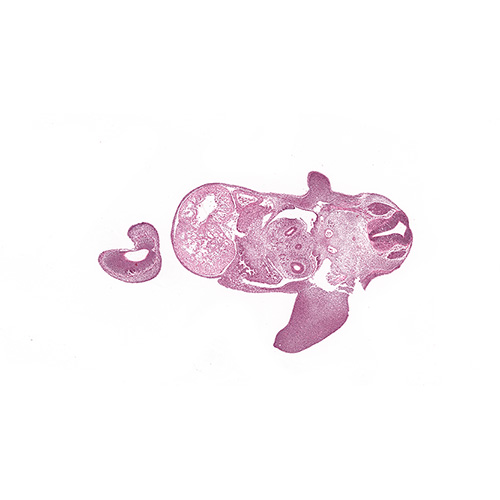 |
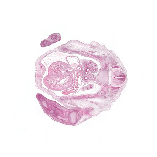
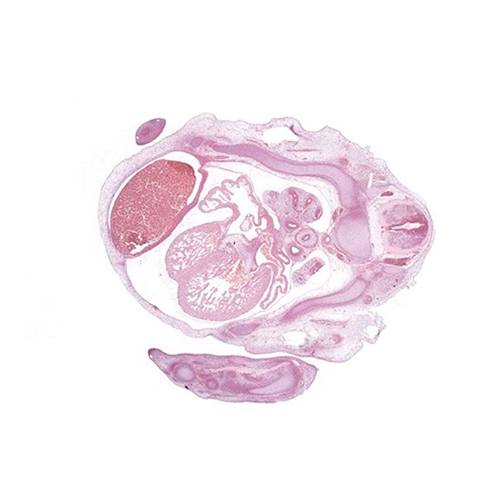
| CS10 | CS11 | CS12 | CS13 | CS14 |
| Transverse | Transverse | Transverse | Transverse | Transverse |
| Longitudinal (Between sagittal and frontal) | Sagittal | Sagittal | Sagittal | Sagittal |
| Frontal | Frontal | Frontal | Frontal | |
 |
 |
 |
 |
 |
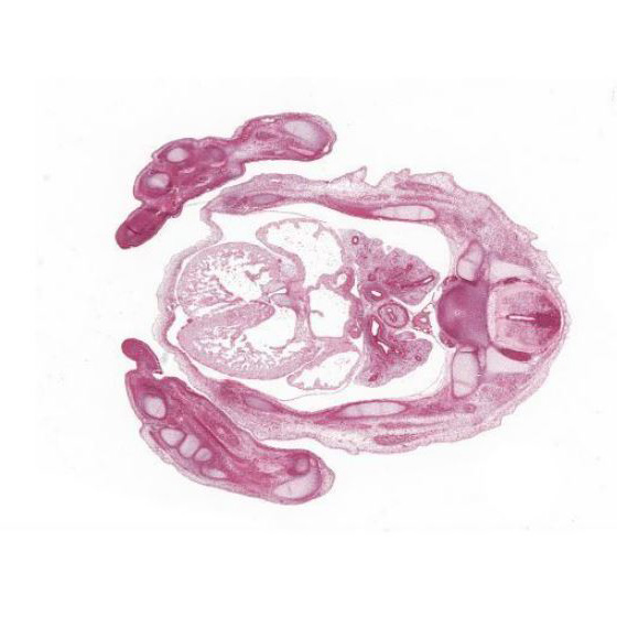
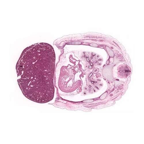
| CS15 | CS16 | CS17 | CS18 | CS19 |
| Transverse | Transverse | Transverse | Transverse | Transverse |
| Sagittal | Sagittal | Sagittal | Sagittal | Sagittal |
| Frontal | Frontal | Frontal | Frontal | Frontal |
 |
 |
 |
 |
|
| CS20 | CS21 | CS22 | CS23 | |
| Transverse | Transverse | Transverse | Transverse | |
| Sagittal | Sagittal | Sagittal | Sagittal | |
| Frontal | Frontal | Frontal | Frontal | |
 |
 |
 |
 |
 |
| Late 8PCW | 9PCW | 10PCW | 11PCW | 14PCW |
| Transverse | Transverse | Transverse | Transverse | Transverse |
| Sagittal | Sagittal | Sagittal | Sagittal | Sagittal |
| Frontal | Frontal | Frontal | Frontal | Frontal |
The eHistology Viewer was developed by Professor Richard Baldock as part of the Edinburgh Mouse Atlas Project
and adapted for the HDBR atlas by Presence Multimedia.
Sections were prepared by Moira Crosier, Lynne Overman, Jasmin Turner, Tamil Dhanaseelan and Steven Lisgo.
Images, Janet Kerwin.